Office of Research & Development |
 |
Talk to the Veterans Crisis Line now
An official website of the United States government

Researchers using stem cells bathed in growth-inducing proteins and grafted onto the injured spinal cords of rats were able to create new cell growth across the injuries and restore some movement to the animals. The scientists, with VA and the University of California, San Diego, reported the advance in the Sept. 14 issue of the journal Cell.
The researchers say the findings may be "the most comprehensive demonstration to date" of the ability of newly formed nerve cells to send messages across a completely severed spinal cord and restore at least partial function.
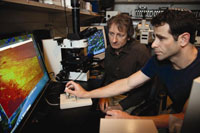
The researchers, led by Paul Lu, PhD, and Mark Tuszynski, MD, PhD, both with VA and UCSD, developed a combination approach to regenerate the injured spinal cord. They used stem cells derived from the nervous system—of both rats and humans, in separate experiments—and embedded the cells in a mesh-like protein called fibrin, part of the body�s blood-clotting system. The fibrin bed contained a "cocktail" of nearly 10 different proteins known to promote nerve-cell growth. The gel-like mixture of stem cells and growth factors was then grafted onto the spinal cords, which had been completed severed, representing the most severe form of spinal cord injury.
"Using this method, after six weeks, the number of axons emerging from the injury site exceeded by 200-fold what had ever been seen before," says Tuszynski. "The axons also grew 10 times the length of axons in any previous study and, importantly, the regeneration of these axons resulted in significant functional improvement."
The human stem cell line used in the experiments is also being used in a clinical trial involving patients with amyotrophic lateral sclerosis, or ALS, at Emory University. The researchers say this suggests a higher potential for "translation" to human therapy.
The work was part of the VA Spinal Cord Injury Collaborative Translational Consortium. The initiative, started in 2010, is building teams of leading scientists—almost a "Who�s Who" of spinal cord research in the U.S. today—to foster high-risk, high-return ideas and to fast-track experimental therapies that may have the potential to reverse spinal cord injury.