Office of Research & Development |
 |
On Jan. 28, 1944, in the midst of World War II, President Franklin D. Roosevelt signed an executive order that stated, in part, that "no blinded servicemen from World War II would be returned to their homes without adequate training to meet the problems of necessity imposed upon them by their blindness."
Today, VA's extensive network of low-vision rehabilitation programs helps many blinded and low-vision Veterans improve their level of functioning. VA's Office of Blind Rehabilitation Services estimates there are approximately 130,000 Veterans in the United States who are legally blind, and more than a million Veterans who have low vision that causes a loss of ability to perform necessary daily activities.
In older Veterans, major causes of vision loss include age-related macular degeneration, glaucoma, cataracts, stroke, and diabetic retinopathy. Among Veterans who have served in Iraq and Afghanistan, blast-related brain injuries can be followed by vision problems such as blurred vision, double vision, sensitivity to light, and difficulty reading. VA estimates that as many as 64 percent of service members with traumatic brain injuries (TBIs) also have a vision problem.
Throughout the nation, VA operates 13 Blind Rehabilitation Centers (BRCs). These are residential inpatient training programs that help Veterans adjust to their blindness. BRCs offer a variety of courses designed to help blinded Veterans achieve a realistic level of independence. VA also operates 22 intermediate and advanced low vision clinics and 23 advanced ambulatory low vision clinics.
The Vision Impairment Services in Outpatient Rehabilitation (VISOR) program provides short-term (about two weeks) blind and vision rehabilitation. They provide overnight accommodations for Veterans and active duty service members who are visually impaired and require lodging. Those who attend VISOR must be able to perform basic activities of daily living independently, including the ability to self-medicate. There are nine VISOR program locations at VA facilities throughout the United States.
The Visual Impairment Center to Optimize Remaining Sight (VICTORS) program complements existing BRCs to support Veterans who are not blind but have significant visual impairment. VICTORS provides rehabilitation through offering definitive medical diagnosis and functional visual evaluation, prescribing low-vision aids and training Veterans in their use, and providing counseling and follow-up. There are currently three VICTORS programs, at Palo Alto, California; Northport, New York; and Lake City, Florida.
Additional information about these programs, and their locations, is available here.
Brain Games
An arguably unsung hero of his time, VA physician and researcher William Oldendorf developed the prototype for the modern-day CT scan. He could not stand seeing his patients suffer through the top medical recommendations at the time for studying brain injury. Today, VA providers at the Cleveland VA, Drs. Walker and Fu bring their passion and professional expertise to the realm of virtual reality as they study another unsung struggle: eye focus issues resulting from mild traumatic brain injuries.
From the invention of the CT scan in the 1960s to today's emerging virtual reality, VA has used every technological innovation it can to understand better, study, and treat traumatic brain injuries, or TBIs.
VA research projects in the area of vision loss and vision restoration cover the entire spectrum of Veterans' needs. In addition to developing vision-restoring treatment, VA investigators are designing and improving assistive devices for those with visual impairments, as well as doing work on a number of innovative wayfinding systems to help Veterans with vision loss navigate in various environments and perform everyday tasks. They are also developing more accurate and efficient methods of vision testing, and are studying the connections between injury and vision loss in eyes that have suffered no overt damage.
VA's Atlanta-based Center for Visual and Neurocognitive Rehabilitation is focused on enhancing Veterans' health by conducting research on the rehabilitation of visual and related neurological impairment, neurocognitive rehabilitation (improving brain function from injury), and retinal and neural repair to prevent or mitigate vision loss resulting from injury and disease. Center researchers are conducting a number of projects to help train blind people and those with low vision to find their way around independently with greater ease. They are also working on projects to provide improved access to eye care for Veterans living in rural regions.
Researchers at the VA Center for the Prevention and Treatment of Visual Loss, located at the Iowa City VA Health Care System, focus on the early detection of potential blinding disorders of Veterans and the general population. These include retinal disease, glaucoma, and TBI. Researchers at the center are evaluating new diagnostic tools that provide increased access to care through telemedicine and the use of automated analysis using portable devices by non-eye care providers, with the goal of providing Veterans with faster access to care and preventing vision loss.
For more on Vision Loss, visit our Prosthetics topic page.
If you are interested in learning about joining a VA-sponsored clinical trial, visit our research study information page.
VA's specialized care for blinded Veterans began with the establishment of the first Center for Rehabilitation of the Blind at the Hines VA Hospital in Chicago in 1948. In this program, selected blind Veterans were trained in a variety of skills.
In the early years after World War II, VA researchers began testing obstacle detectors built for the U.S. Army during the war to find ways to use them to help blinded Veterans navigate their environment. These obstacle detectors eventually led to the development of the laser cane, still in use today. Laser canes emit pulses of infrared light and make different sounds to indicate obstacles ahead, or drop-offs such as a street curb.
In the 1950s, '60s, and '70s, VA researchers and research funding helped develop devices that produced speech-like sounds in response to letter shapes. These "reading machines" were the predecessors for technologies now in widespread use.
Comparative effectiveness of intraocular lenses for cataract surgery and lens replacement—Cataract is an eye condition in which the natural crystalline lens becomes cloudy and can ultimately lead to poor vision. It is estimated that half of all Americans older than age 75 have cataract or have had cataract surgery. The surgical removal of cataracts and the implantation of prosthetic lenses is one of the most common surgeries performed in the United States.
In a comparative-effectiveness study published in 2017, VA researchers reviewed previous studies on two types of lenses commonly used in cataract surgery: monofocal lenses, which promote clear distance vision but require reading glasses, and multifocal lenses, which produce multiple focal points and allow a full range of vision—near, far, and in-between.
They found that multifocal lenses help people to see without glasses and improve the clarity and sharpness of vision for nearby objects, such as books, without sacrificing distance vision; and that they result in better visual function and quality of life than monofocal lenses. However, they also result in poorer contrast sensitivity and create a greater risk of developing glare and halos. The researchers found “low strength” evidence that more people are dissatisfied with monofocal lenses than multifocal ones.
The team also noted that none of the studies they reviewed were performed on Veterans receiving care from VA, or even on Americans, so the applicability of their results to VA patients is uncertain. They suggested VA conduct a multisite randomized clinical trial to provide higher quality evidence than currently exists about the benefits, harms, necessary resources, and costs of the two procedures.
Advances in body armor, medical care, and equipment have enabled more service members to survive devastating blast injuries. Use of polycarbonate eye protection has reduced the number of open-globe eye wounds service members sustain, but this eye protection does not guard against the effect of blast waves. Researchers at the VA Palo Alto Health Care System have completed a number of studies related to the effects of blasts on vision.
Sensory issues—Sensory problems are common among Veterans who have had TBIs. In 2012, Palo Alto researchers reported on a study of 21,000 Veterans evaluated for TBI in VA outpatient clinics. They found that 9.9 percent of them reported vision problems, 31.3 percent reported hearing impairments, and 34.6 percent reported both vision and hearing issues.
Another study by the team, published in 2013, looked at 50 Veterans with blast-related TBI. They found that more than 65 percent had vision problems, and 77 percent reported sensitivity to light. Other difficulties included problems with eye movement, blurred vision, and problems focusing. The same number of Veterans with non-blast-related TBI was also studied, and the rates of vision complaints were similar.
In 2016, this team published an additional study indicating that perimetry (visual field testing) for Veterans with TBIs within two months of their combat blast exposure provides a reliable indicator of long-lasting vision problems. These tests also reveal high rates of visual-field deficits among those tested, indicating that blast wave forces may significantly affect both the eye and visual pathways.
Hidden eye injuries also a problem—Another Palo Alto study, published as a letter to the editor in the New England Journal of Medicine in 2011, found that many Veterans who have had blast injuries also have "hidden eye injuries" that may go undetected without comprehensive eye examinations.
The research team evaluated 46 combat Veterans, 43 men and three women, who had developed TBIs from documented blast injuries, and had not previously reported any injury to their eyes. Of that group, 20 (43 percent) suffered what ophthalmologists call "closed eye ocular injuries"—injuries that do not actually penetrate the eye.
The types of injuries the team found included damage to the cornea, the iris, and the optic nerve. They also found injuries to other areas of the eye as well. Accordingly, the researchers recommended that any patient with a TBI diagnosis, including those without current vision problems, have a comprehensive eye examination by an ophthalmologist to check for hidden eye injuries that may cause future problems such as glaucoma.
As a result, the Vision Center of Excellence, operated jointly by VA and the Department of Defense (DoD), has provided clinical recommendations to eye care professionals, especially VA and DoD providers, to guide optometrists and ophthalmologists in treating patients with eye and vision problems following a possible TBI or blast exposure. The recommendations include tests to help eye care providers screen and assess for the most common ocular injuries as well as vision problems caused by or associated with TBI.
The Palo Alto research team led a larger team of VA researchers that developed, in 2013, a clinical tool that can be used by eye-care providers either as a screening tool or as part of a full eye examination when they see a patient with a history of having had a mild TBI. The tool includes guidance for collecting a history of the patient's experiences, and for measuring his or her vision function. This tool is included in the Vision Center of Excellence’s clinical recommendations, and is now being used throughout VA and DoD.
According to a report by VA's Office of the Inspector General (IG), cataract surgery is one of the most common surgeries performed in VA. Because most VA facilities are teaching hospitals, many cataract surgeries are performed by ophthalmologic residents. (Residents are medical-school graduates engaged in specialized practice under supervision in a hospital.) VA regulations require residents conducting cataract surgery to be directly supervised by an attending ophthalmologist who must be physically present in the operating room, and the IG report found that VA facilities generally complied with this requirement.
A 2016 study led by researchers with the VA Boston Health Care System looked at more than 4,200 cataract surgery cases at VA facilities throughout the nation, and found that Veterans who were operated on by residents had an overall significant improvement in visual acuity (the clarity of their vision) and visual function (the ability to discern forms, colors, and movement) compared with before their surgery, even if complications arose as a result of their procedure. Those who had complications, however, showed a less marked improvement in their vision.
Precision navigation support—The Atlanta VA Medical Center, with funds from the National Institutes of Health, is working to develop a precision navigation application for people without sight. Existing navigation apps rely on GPS technology, which can typically guide people to within sighting distance of a location but cannot provide the cane-reach accuracy that is needed by those without site. Researchers are analyzing the unique walking patterns of people who use a white cane to accurately track their step-by-step location, such as a building exit. When combined with GPS data, the team hopes, the application will achieve cane-reach accuracy.
The research team is also developing a mapping database whereby satellite maps can be annotated to indicate the exact coordinates of building entrances, crosswalks, bus stops, and other such locations to allow the app to guide the user to within cane-touch distance of known locations.
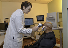
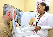
Technology-based Eye Care Services helps homeless and rural Veterans—The Atlanta VA Medical Center’s eye clinic has developed an eye-screening program called Technology-based Eye Care Services. The program, launched in 2015, allows patients to be checked for eyeglasses and screened for common eye diseases using special equipment and eye photographs at their local VA community-based outpatient clinic. In a 2017 study of the program, researchers found the program significantly improved access to eye care at the facility, reduced health care costs, and provided care to a significant percentage of homeless Veterans. The researchers recommended that other health care facilities consider using a similar protocol to extend care to at-risk patients.
Veterans Innovative Sight in Optometry Nexus program—V.I.S.I.O.N. is a project that aims to improve access to eye care, prevent blindness, and provide Veterans with vital eye care services. The platform for the program is a high-efficiency technology suite created in the old eye examination space in the White River Junction VA Medical Center, Vermont.
The suite is filled with the most efficient equipment available on the market, and optometrists and other staff use the new technologies in the room to carry out nearly 10 tests that could not previously fit in one area. In addition to taking up little space, the equipment provides highly accurate information and eliminates the need to move a patient to three or four rooms to obtain the same results. The facility hopes the program will be expanded to other VA facilities throughout the nation.
Antioxidants may prevent vision loss from SLOS—A 2018 study led by researchers at the VA Western New York Healthcare System, the Louis Stokes VA Medical Center in Cleveland, Case Western Reserve University, and the University of Buffalo has demonstrated that the addition of widely available antioxidants to the current standard of care can prevent vision loss in an animal model of Smith-Lemil-Opitz Syndrome (SLOS), a rare genetic disease.
Caused by the body’s inability to make cholesterol, the disease is a birth defect that results in multiple sensory and cognitive abnormalities, physical deformities, and disabilities including vision loss. The team found that oxidation of a specific molecule was the key to the disease’s mechanism, and correctly hypothesized that blocking the oxidation with antioxidants should prevent or significantly reduce the severe retinal degeneration observed in rodents.
The team hopes to conduct additional animal model studies, and then investigate whether the treatment will work in humans with SLOS.
Chemical reaction in the eye may improve vision—In a 2017 study led by researchers at the Louis Stokes VA Medical Center in Cleveland and Case Western Reserve University, the team found that a light-sensing pigment that is found in everything from bacteria to vertebrates can be biochemically manipulated to reset itself.
Researchers used a modified form of vitamin A, called locked retinal, to induce the recycling mechanism and engage proteins called retinals that are central to human vision. The discovery will help scientists better understand the biochemistry of vision and why the chemical configuration of retinals is critical in enabling humans to receive light. Their discovery opens an opportunity to modify retinals’ functioning to help improve vision.
Stem cells can regenerate lenses after cataract surgery—Researchers at VA and the University of California, San Diego, and colleagues in China, have developed a regenerative medicine approach to remove congenital cataracts in infants, allowing stem cells to regrow functional lenses. The approach was described in a 2016 paper.
Congenital cataracts involves lens clouding at birth or shortly thereafter, and is a significant cause of blindness in children. The clouded lens obstructs the passage of light to the retina and visual information to the brain, resulting in significant visual impairment. The new treatment, a surgical technique that stimulates epithelial stem cells in the lens, produced fewer surgical complications than the current standard of care and resulted in regenerated lenses with superior visual function in all 12 of the children under age 2 who received the new surgery. The team hopes to expand their work to treating age-related cataracts, the leading cause of blindness in the world.
Interdisciplinary low-vision services not better than basic low-vision services for patients with mild macular degeneration—A team of VA researchers completed a low-vision intervention trial (LOVIT II) in 2016 to determine if an interdisciplinary low-vision rehabilitation program is more effective than basic low-vision care provided by an optometrist working alone to improve visual reading ability in Veterans with macular diseases.
Interdisciplinary services were provided by both optometrists and low-vision therapists, and included therapy to improve the use of patients’ remaining vision and low-vision devices, and structured homework to practice the use of low-vision devices that are prescribed and dispensed.
The team found that both methods of care were effective, but the interdisciplinary services were more effective only for patients with significant visual impairment. Basic services, they found, may be sufficient for most low-vision patients with mild impairment.
Comparative effectiveness of anti-vascular endothelial growth factor agents—Among the diseases commonly responsible for substantial vision loss in adults are age-related macular degeneration, diabetic macular edema, and central or branch retinal vein occlusion. All of these diseases are driven at least in part by vascular endothelial growth factors (VEGFs). Several drugs called anti-VEGF agents block these factors and limit eye damages. Some can slow and even reverse the vision loss typically seen in patients with these diseases.
Investigators with VA’s Evidence-based Synthesis Program Center in Portland, Oregon, conducted a systematic review of existing data sources, completed in 2017, to compare the effectiveness, harms, and costs of commonly used anti-VEGF agents. They found no clear, consistent, clinically meaningful differences between the drugs for most outcomes. The drug aflibercept may provide a greater short-term visual acuity benefit than other agents among patients with low visual acuity, but longer-term findings are unclear. The drug bevacizumab appeared to be significantly more cost-effective than the other drugs in its compounded form.
The team concluded that in choosing among available drugs, clinicians should consider factors such as patient preferences; individual treatment responses; convenience; and the distance the patient has to travel to the treatment facility, because some forms of treatment require more visits to health care facilities than others.
Exercise protects against retinal degeneration—A 2014 study by researchers at the Atlanta VA Medical Center and Emory University suggested that physical activity can protect eyes as they age.
The researchers ran mice on a treadmill for two weeks before and after exposing the animals to bright light that causes retinal degeneration. They found that treadmill training preserved photoreceptors and retinal cell function in the mice. The exercised animals lost only half the number of photoreceptor cells as animals that spent the same amount of time on a stationary treadmill.
The researchers believe that their work may one day lead to tailored exercise regimens or combination therapies in treatment of retinal degenerative diseases.
In a 2017 study, the research team found that increased physical activity was associated with greater self-reported visual function and improved quality of life in people with retinitis pigmentosa.
Eye vitamins may not be valuable—In 2001, a landmark non-VA study, the Age-Related Eye Disease Study (AREDS), found that a specific formula of supplements containing high doses of zinc and other antioxidants could slow the deterioration of the eye's macula (the central part of the retina). Age-related macular degeneration (AMD) is the leading cause of blindness among older adults.
A follow-up study called AREDS2, conducted in 2011, found that the formula remained effective even if one ingredient, beta-carotene, was replaced with related nutrients. Both studies offered formulas that could be used to prepare a compound that might protect against AMD. As a result, a number of vitamin manufacturers have begun to sell ocular nutritional supplements, designed to keep those who use them from developing AMD.
In 2016, a research team from the Providence VA Medical Center and three universities examined 11 different supplements to see if they were prepared in accordance with either the ARED or ARED2 formula.
The team found that while all of the supplements contained some ingredients from either formula, only four of the products contained doses equivalent to those in the study.
Another four products contained lower doses, and four included additional vitamins, minerals, or herbal extracts that were not part of the original studies. None of the supplements precisely duplicated either the AREDS or AREDS2 formulas.
The team concluded that without clinical research, it is impossible to determine how those additional ingredients affect the formula, and that ophthalmologists should educate their patients on what to look for in supplements.
Lowering high blood pressure reduces vision risk—The Durham VA Medical Center-based Hypertension Intervention Nurse Telemedicine Study looked at the effects of telephone-based medication management and behavioral management on high blood pressure in people with diabetes.
In 2013, the study team found that patients whose blood pressure improved due to the intervention also had about half the risk of worsening diabetic retinopathy, compared with those receiving usual care. Diabetic retinopathy damages the retina, and can eventually lead to blindness.
Cost-effectiveness of teleretinal screening—VA researchers have also looked at the department's existing teleretinal screening process, in which special cameras take images of the lining inside the eye. They have found that telemedicine is a cost-effective way to screen Veterans less than 80 years of age for signs of diabetic retinopathy, a complication of diabetes that affects the eyes.
A 2013 VA study found, however, that it is not cost-effective to use telemedicine for diabetic retinopathy screening in patients aged 80 or older, or for populations of under 3,500 patients.
The SightBook smartphone app allows patients to test their vision frequently on their smartphones and share the test results with their designated physician in real time.
Tests available on the app address visual acuity; visual disturbances caused by changes in the retina; visual acuity at low light; contrast sensitivity (the ability to see objects that may not be outlined clearly); and several other aspects of vision health.
In 2016, a research team from the Miami VA Healthcare System and the University of Miami tested the accuracy of readings from SightBook compared with readings using a Snellen eye chart, the standard eye chart that is read at a distance of 20 feet (or with mirrors that make it appear to be at a distance of 20 feet); and with near card eye charts, which are designed to be read at shorter distances.
They found that while there were discrepancies in results between each of the methods of testing visual acuity, the results from each method could be successfully reproduced, and that baseline SightBook acuity measures allow for future vision comparisons.
VA employees showcase innovative projects, best practices aimed at helping Vets, VA Research Currents, Aug. 17, 2017
Some top-selling eye vitamins don't match scientific evidence, says study, VA Research Currents, Jan. 21, 2015
Helping those with vision loss find their way, VA Research Currents, Oct. 21, 2014
Lab study: Exercise wards off retinal damage, VA Research Currents, March 26, 2014
Video: Vision Loss Research
VA Center for the Prevention and Treatment of Visual Loss
Center for Visual and Neurocognitive Rehabilitation
Prevention of retinal degeneration in a rat model of Smith-Lemil-Opitz syndrome. Fliesler SJ, Peachey NS, Herron J, Hines KM, Weinstock NI, Ramachandra Rao S, Xu L. Combining dietary supplementation with cholesterol synthesis can better preserve retinal structure and function in patients with this syndrome than current standards of care. Sci Rep. 2018 Jan 19:8(1):1286.
Physical activity and quality of life in retinitis pigmentosa. Levinson JD, Joseph E, Ward LA, Nocera JR, Perdue MT, Bruce BB, Yan J. In retinitis pigmentosa, increased physical activity is associated with greater self-reported visual function and quality of life. J Ophthalmol. 2017;2017:6950642. (Epub ahead of print)
Cataract surgery practices in the United States Veterans Health Administration. Havnaer AG, Greenberg PB, Cockerham GC, Clark MA, Chomsky A. In cataract surgery in the VHA, routine preoperative testing is commonly performed, and emerging practices have limited roles. J Cataract Refract Surg. 2017 Apr;43(4)543-551.
Photocyclic behavior of rhodopsin induced by an atypical isomerization mechanism. Gulati S, Jastrzebska B, Banerjee S, Placeres AL, Miszla P, Gao S, Gunderson K, Tochtrop GP, Filipek S, Katayama K, Kiser PD, Mogi M, Stewart PL, Palczewski K. A light-sensing pigment found in everything from bacteria to vertebrates can be biochemically manipulated to reset itself. Proc Natl Acad Sci U S A. 2017 Mar 28;114(13):E2608-E2615.
Remote eye care screening for rural Veterans with Technology-based Eye Care Services: a quality improvement project. Maa AY, Wojciechowski B, Hunt K, Dismuke C, Janjua R, Lynch MG. A quality improvement project at the Atlanta VA Medical Center to provide eye screening services improved access to care, provided care to homeless Veterans, and reduced health care costs. Rural Remote Health. 2017 Jan-Mar;17(1):4045.
Eye care productivity and access in the Veterans Affairs health care system. Lynch MG, Maa A, Delaune W, Chasan J, Cockerham GC. Eye care technicians provide a cost-effective multiplier effect for provider productivity, especially in ophthalmology clinics. Mil Med. 2017 Jan;182(1):e1631-e1635.
Comparative clinical and economic effectiveness of anti-vascular endothelial growth factor agents (internet). Low A, Lansagara D, Freeman M, Fu R, Bhavsar K, Faridi A, Kondo K, Paynter R. The most commonly used anti-vascular endothelial growth factor drugs have been shown to slow and even reverse vision loss. This report compares the effectiveness, harms, and costs of these drugs. VA Evidence-based Synthesis Program Report. 2017 Jan.
Outcomes of the Veterans Affairs low vision intervention trial II (LOVIT II): a randomized clinical trial. Stelmack JA, Tang XC, Wei Y, Wilcox DT, Morant T, Brahm K, Sayers S, Massof RW; LOVIT II Study Group. Basic low vision services may be sufficient for most low vision patients with mild visual impairment. JAMA Ophthalmol. 2016 Dec. 15 (Epub ahead of print).
Reproducibility and comparison of visual acuity obtained with SightBook mobile application to near card and Snellen chart. Phung L, Gregori NZ, Ortiz A, Shi W, Schiffman JC. The SightBook mobile app offers a new portable vision assessment tool for the office and remote patient monitoring. Retina. 2016 May;36(5):1009-20.
Ophthalmic complications related to chemotherapy in medically complex patients. Harman LE. Recognizing potentially serious ocular complications of cancer therapy before they result in irreversible injury starts with taking a relevant clinical history and performing a basic eye examination. Cancer Control. 2016 Apr:23(2):150-6.
The use of telemedicine to extend ophthalmology care. Lynch MG, Maa AY. Teleopthalmology is a valuable tool for extending care to larger populations of high-risk patients, and can be a valuable care extender in places where emergency departments do not have an ophthalmologist on call. JAMA Ophhalmol. 2016 Mar 24. (Epub ahead of print)
Lens regeneration using endogenous stem cells with gain of visual function. Lin H, et al. A new regenerative medicine approach can remove congenital cataracts in infants, permitting remaining stem cells to regrow functional lenses. Nature. 2016 Mar 17;531(7594):323-8.
Outcomes of cataract surgery with residents as primary surgeons in the Veterans Affairs Healthcare System. Payal AR, Gonzalez-Gonzalez LA, Chen X, Cakiner-Egilmez T, Chomsky A, Baze E, Vollman D, Lawrence MG, Daly MK. Resident-operated cases with and without events had an overall significant improvement in visual acuity and visual function compared with preoperatively. J Cataract Refract Surg. 2016 Mar;42(3):370-84.
Automated perimetry and visual dysfunction in blast-related traumatic brain injury. Lemke S, Cockerham GC, Glynn-Milley C, Lin R, Cockerham KP. Reliable automated perimetry can be accomplished in most patients with TBI from combat blast exposure and reveals high rates of visual field deficits, indicating that blast forces may significantly affect the eye and visual pathways. Ophthalmology. 2016 Feb;123(2):415-24.
Corneal mechanical thresholds negatively associate with dry eye and ocular pain symptoms. Spierer O, Felix ER, McClellan AL, Parel JM, Gonzalez A, Feuer WJ, Sarantopoulos CD, Levitt RC, Ehrmann K, Galor A. Mechanical detection and pain thresholds measured on the cornea are correlated with dry eye symptoms and ocular pain. Invest Ophthalmol Vis SCi. 2016 Feb;57(2):617-25.

Download PDF
 Researchers take new approach to detect, treat eye disease that can lead to blindness
 Research improves tele-eye screening for Veterans